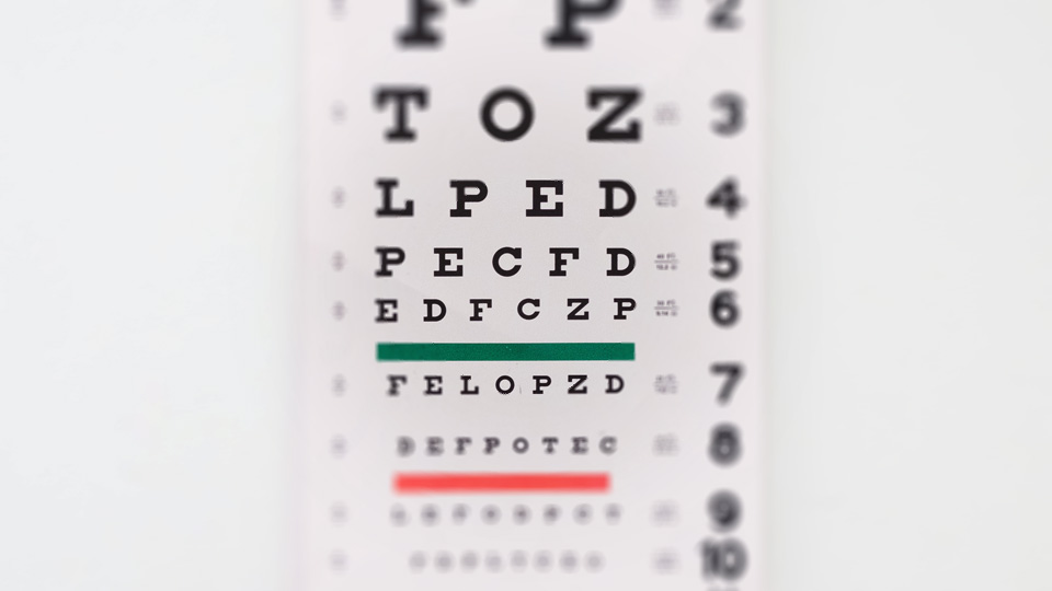
Comprehensive Eye Examination

A complete eye examination is administered by Dr Gerard Nah and his team. This examination aims to exclude common eye diseases like binocular vision problems, cataract, glaucoma, retinal tears and holes, etc.
This appointment will take at least:
- 2 hours for adults
- 3 hours for children aged 12 and below
There are 3 main steps in this examination.
-
Refraction
It is the process of determining the eye's refractive power. During refraction, the optometrist puts a trial frame with lenses in front of your eyes to show you a series of choices. By going through this process, the optometrist will be able to accurately measure your spectacle prescription. This is a necessary step in establishing the functional status of your eye.
-
Dilated fundus examination
Dilating drops are instilled in your eyes to enlarge the pupils, allowing a better view of the internal structures of the eye (retina). The pupil is like a window that expands and contracts controlling the amount of light entering the eye. By opening the pupil, the inside of your eye can be better visualized.
-
Slit lamp examination
A slit lamp is a special microscope which provides a narrow beam of light which helps the doctor examine of the various structures of the eye.
Other relevant tests may then be performed depending on your concerns and results of the above-tests on the day of your examination.
Eye screening is important as it helps to detect potentially sight-threatening eye conditions such as cataract, glaucoma, age-related macular degeneration, diabetic retinopathy and refractive errors. It is recommended that eye screening is conducted around the age of 40, or earlier if there is a family history of eye disease.
Where required, your ophthalmologist will order other specialized tests. The following are examples of such tests.
- Binocular Ophthalmoscopy of the retina
- Fluorescein staining
- Amsler Grid testing
- Visual Fields by Confrontation
- Goldmann/Tonopen Tonometry
- Schirmer's Tear Test (STT)
- Gonioscopy of the angles of the eyes
- Automated Visual field tests
- Topography/Tomography of the Cornea
- Tear Film Assessment
- Scheimpflug Tomography of the Cornea and Anterior Segment of the eye
- Optical Coherence Tomography (OCT) of the Cornea, Anterior Chamber and Lens
- Optical Coherence Tomography (OCT) of the Macular and Optic Nerve
- Ultra Wide Field Laser Scanning Ophthalmoscopy of the Retina
- Anterior Segment Analysis and/or Photography
- Colour Vision testing
- Stereopsis (3D) testing
- Contrast Sensitivity Testing
- Ocular Motility Testing and Measurement of latent and manifest squints
- Ultrasound scans of the eye
- Ultrasound Biometry of the eye
- Optical Biometry of the eye
- Fluorescein and Indocyanine Green (ICG) Angiography of the Retina and Choroid